Finding a sternum lump in the middle of your chest can be an alarming experience, particularly for women who immediately worry about underlying conditions such as breast cancer or cardiac anomalies. In 2026, clinical diagnostics and imaging technologies allow healthcare providers to differentiate between benign musculoskeletal variants and more serious pathological growths with high precision. This comprehensive guide explores the anatomical structures of the sternum, the common and rare differential diagnoses for a central chest lump in females, diagnostic protocols, and when to seek immediate medical intervention.
Anatomical Context and Structural Overview of the Female Sternum
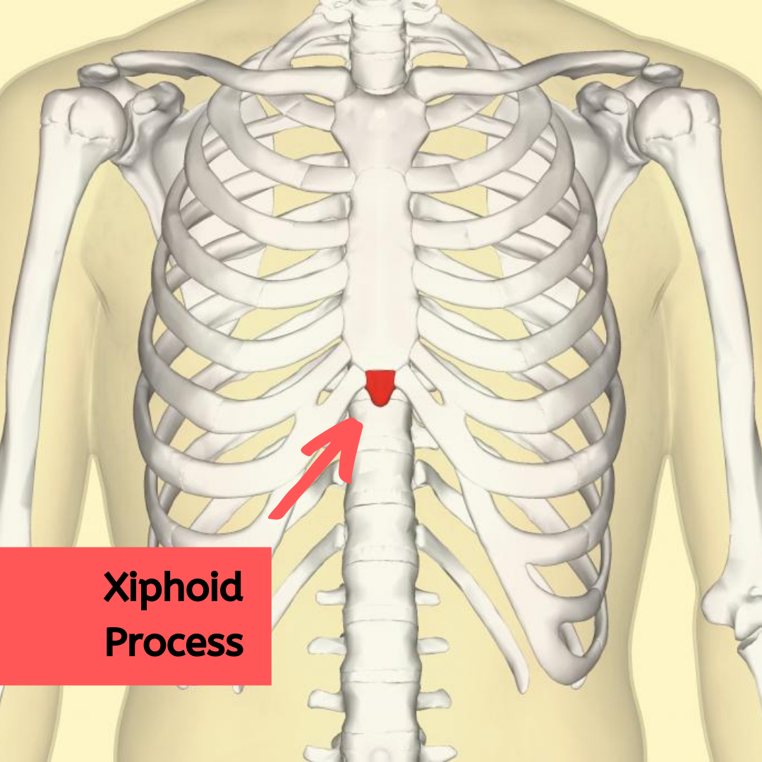
The sternum, commonly known as the breastbone, is a flat bone situated at the anterior center of the chest. It consists of three primary segments: the manubrium at the superior end, the body (gladiolus) in the middle, and the xiphoid process at the inferior tip. In females, the overlaying soft tissues, including pectoral muscles, breast tissue, and subcutaneous fat, can sometimes obscure or accentuate variations in this bony architecture.
When a woman detects a lump directly over the sternum, the mass can originate from skin structures, subcutaneous tissue, the cartilage connecting the ribs to the breastbone, or the bone itself. Understanding the exact location along the manubrium, body, or xiphoid process helps clinicians narrow down the potential causes during an initial physical examination.
Differential Diagnoses: Benign Versus Pathological Causes
A thorough clinical evaluation classifies sternal masses into various categories based on tissue origin, consistency, and mobility.
- Costochondritis and Tietze Syndrome: Inflammation of the costal cartilages connecting the ribs to the sternum. While often presenting as sharp chest wall pain, Tietze syndrome specifically causes a distinct, localized, non-pitting swelling—frequently near the second or third rib cartilage—that can mimic a true bony lump.
- Xiphoid Process Variations: The xiphoid process varies significantly among individuals. It can be bifid, angled sharply forward, or enlarged, frequently leading patients to mistake this normal anatomical feature for an abnormal growth.
- Lipomas and Epidermoid Cysts: Benign soft-tissue tumors. Lipomas are slow-growing, movable fatty lumps under the skin, while epidermoid cysts are dermal nodules containing keratin that may occasionally become tender or inflamed.
- Chondromas and Osteochondromas: Benign bone and cartilage tumors that develop on the sternum. Though rare, these slow-growing lesions can present as hard, fixed masses attached directly to the breastbone.
- Primary or Metastatic Malignancies: Rare malignant tumors such as chondrosarcoma, multiple myeloma, or metastatic lesions from primary sites (such as the breast or lung) can manifest as firm, fixed, and occasionally painful sternal masses.
Clinical Observation Notice
Benign soft tissue growths like lipomas are typically mobile beneath the skin and non-tender, whereas primary bone tumors or malignant lesions tend to feel hard, immovable, and firmly anchored to the underlying sternal periosteum. Any fixed, hard, or rapidly changing mass warrants immediate professional assessment.
Comparative Analysis of Common Central Chest Masses
To help contextualize the physical characteristics of various sternal and chest wall abnormalities, the following comparative matrix outlines their clinical presentations, typical locations, and general management strategies.
| Condition Type | Typical Location | Physical Characteristics | Common Patient Age/Demographic | Standard Clinical Management |
|---|---|---|---|---|
| Xiphoid Process Prominence | Inferior sternum tip | Hard, fixed, non-tender, matches bone contour | Any age (often noticed during weight loss) | Reassurance and observation |
| Tietze Syndrome | Costochondral junctions (2nd/3rd ribs) | Localized, firm, tender swelling | Females aged 20-40 | NSAIDs, physical therapy, localized heat |
| Subcutaneous Lipoma | Overlying soft tissue | Soft, mobile, rubbery, painless | Adults over 30 | Observation or simple excision if symptomatic |
| Epidermoid Cyst | Dermal layer of chest skin | Firm, punctum often visible, movable | Adults | Warm compresses, minor surgical removal if infected |
| Osteochondroma | Sternal body or manubrium | Hard, immobile, painless or dull ache | Young adults to middle-aged | Imaging, potential surgical resection if growth occurs |
| Sternal Malignancy | Any part of sternum | Hard, fixed, painful, rapid growth | Older adults or high-risk history | Oncology referral, biopsy, advanced staging |
Diagnostic Workup and Imaging Protocols in 2026
When a female patient presents with a mid-chest sternal lump, healthcare providers follow a systematic diagnostic pathway to rule out malignancy and identify the exact etiology.
- Detailed Clinical History and Physical Palpation: The clinician evaluates the duration of the lump, presence of pain, relation to movement or breathing, and personal or family history of cancer. Palpation determines if the mass is superficial, subcutaneous, cartilaginous, or osseous.
- Diagnostic Radiography (X-rays): Frontal and lateral chest X-rays provide an initial look at the sternal cortex, checking for lytic or blastic bone lesions, cortical expansion, or fractures.
- High-Resolution Ultrasound: Particularly useful for female patients, ultrasound differentiates between fluid-filled cysts, solid lipomas, and cartilaginous swellings without exposing the patient to ionizing radiation.
- Cross-Sectional Imaging (CT or MRI): If ultrasound and X-rays are inconclusive, a computed tomography (CT) scan offers detailed bone architecture assessment, while magnetic resonance imaging (MRI) provides superior soft-tissue contrast for evaluating cartilage, bone marrow, and surrounding musculature.
- Core Needle or Surgical Biopsy: If imaging suggests a primary bone tumor, metastasis, or atypical growth, a tissue sample is harvested for histopathological analysis to establish a definitive diagnosis.
Red Flag Symptoms Requiring Urgent Medical Evaluation
While the vast majority of sternal lumps in females are benign musculoskeletal variants or harmless lipomas, certain symptoms necessitate immediate evaluation by an emergency physician or specialist:
- Rapid and noticeable increase in the size of the lump over a period of weeks.
- Severe, persistent, or worsening pain that radiates to the back, shoulders, or arms, or occurs independently of movement and pressure.
- Skin changes over the lump, including ulceration, redness, dimpling, or fixated tethering of the skin.
- Associated systemic symptoms such as unexplained weight loss, chronic fatigue, night sweats, or persistent low-grade fevers.
- Palpable lymphadenopathy in the axillary (underarm), supraclavicular, or cervical regions.
Frequently Asked Questions
What causes a hard lump in the middle of the female chest?
A hard lump in the middle of the chest is frequently a normal anatomical variant such as a prominent xiphoid process, or a benign condition like costochondritis, but it can occasionally represent a bone tumor or cyst. Professional clinical imaging is required to confirm the exact tissue origin.
Can breast cancer present as a lump on the sternum?
Breast cancer typically develops within the breast tissue itself, but advanced or locally recurrent disease can occasionally involve the chest wall, pectoral muscles, or sternum. Any persistent mid-chest mass in a woman with a personal history of breast cancer must be evaluated promptly by her oncology team.
Is a painful sternum lump more dangerous than a painless one?
Pain is not a reliable indicator of malignancy; many benign inflammatory conditions like Tietze syndrome cause significant pain and tenderness, whereas some slow-growing benign tumors are completely painless. Both painful and painless persistent lumps require professional diagnostic workup.
How do doctors differentiate between a fat lump and a bone lump on the breastbone?
Physicians use physical examination techniques to test mobility and texture, followed by diagnostic imaging such as ultrasound for soft tissue masses like lipomas, and X-rays or CT scans for hard masses anchored to the bone.
When should I schedule an appointment for a chest wall lump?
You should schedule an evaluation with your primary care physician or a specialist if the lump persists for more than two weeks, increases in size, becomes increasingly painful, or is accompanied by systemic symptoms like unexplained weight loss.
Professional Medical Consultation and Next Steps
Discovering any unfamiliar mass on your body warrants a calm, methodical approach. If you notice a sternum lump in the middle of your chest, schedule an appointment with your primary care provider or a women's health specialist for a comprehensive clinical assessment. Early evaluation ensures accurate diagnosis, rules out serious pathology, and provides targeted treatment or comforting reassurance tailored to your specific anatomical health profile.