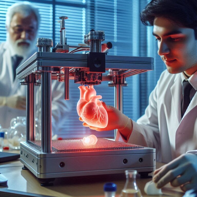
The 3D printed human heart represents a paradigm shift in cardiovascular medicine, transitioning from rigid mechanical devices to patient-specific biological structures. By leveraging advanced bio-printing techniques, medical researchers are laying the groundwork to manufacture functional heart tissue, vascular networks, and ultimately whole organs capable of replacing failing hearts and eliminating the organ donor shortage.
Introduction to Bioprinting in Cardiac Care
For decades, the field of cardiology has battled a fundamental limitation: the human heart possesses a severely restricted capacity for self-repair. When myocardial tissue suffers a catastrophic insult, such as a severe myocardial infarction, the resulting scar tissue cannot contract or conduct electrical impulses effectively. While heart transplantation remains the gold standard for end-stage heart failure, the global shortage of donor organs means that thousands of patients die on waiting lists every year. This reality has catalyzed an extraordinary convergence of bioengineering, stem cell biology, and additive manufacturing.
The concept of the 3D printed human heart moves far beyond industrial plastics and metallic prototypes. Researchers are now working with bio-inks—formulations composed of living human cells suspended in hydrogels derived from extracellular matrices. These biological inks are extruded layer-by-layer through precision nozzles to build intricate three-dimensional structures that mimic the native architecture of the human heart. As this technology matures, it promises to redefine surgical planning, drug discovery, and regenerative medicine, altering the landscape of healthcare for generations to come.
Understanding the Core Technology: How Bio-Printing Works
To appreciate the future of cardiac innovation, one must first understand the mechanics of 3D bioprinting. Unlike traditional 3D printing, which uses melted thermoplastics or photopolymers, bioprinting must protect the viability of living cells throughout the fabrication process.
The process begins with medical imaging data, typically derived from high-resolution magnetic resonance imaging (MRI) or computed tomography (CT) scans of a patient's chest. These scans are converted into computer-aided design (CAD) models, which dictate the precise coordinates for the printhead. The printer deposits bio-inks—often containing induced pluripotent stem cells (iPSCs)—alongside sacrificial support materials. These support baths allow fragile, complex geometries, such as branching coronary blood vessels and intricate chamber walls, to maintain their shape without collapsing under gravity.
- Patient Medical Imaging (MRI/CT)
- CAD Model Generation
- Bio-Ink Preparation (iPSCs + Hydrogel)
- Precision Layer-by-Layer Extrusion
- Cell Maturation & Vascularization in Bioreactor
Once printed, the construct cannot immediately be implanted. It must be transferred to a specialized bioreactor, an environment that simulates physiological conditions like temperature, pH, mechanical stretch, and fluid perfusion. Inside the bioreactor, the cells undergo maturation, forming tight intercellular junctions, aligning themselves into functional muscle fibers, and establishing primitive capillary networks.
Current State of Research and Milestones
While a fully functional, transplantable whole human heart suitable for routine clinical use is still undergoing rigorous preclinical evaluation, significant milestones have already been achieved by leading academic and clinical research institutions worldwide.
In recent years, researchers at institutions such as Tel Aviv University demonstrated the ability to print miniature vascularized hearts using cellular material taken directly from the patient. These mini-hearts possessed cellular, structural, and biochemical properties matching the patient's own biology, reducing the risk of immune rejection. Similarly, teams at the Wyss Institute at Harvard University and Carnegie Mellon University have pioneered advanced embedding techniques, such as Freeform Reversible Embedding of Suspended Hydrogels (FRESH), allowing soft materials like collagen to be printed into complex anatomical shapes.
Major Technical Milestones in Cardiac Bioprinting
| Milestone / Achievement | Description | Primary Significance |
|---|---|---|
| Patient-Specific Micro-Hearts | Printing miniature hearts containing blood vessels, ventricles, and chambers using personal cellular biopsies. | Proves that personalized, immunologically compatible cardiac tissue can be fabricated. |
| FRESH Printing Technique | Embedding soft hydrogels and collagen within a supportive gel bath during extrusion. | Solves the structural collapse problem associated with printing high-moisture biological materials. |
| Vascular Network Integration | Creating branching capillary structures capable of delivering oxygen and nutrients deep within tissue. | Overcomes the diffusion limit, preventing core cell death in thicker tissue constructs. |
| Electrical Synchronization | Inducing printed cardiac muscle cells to beat synchronously in response to electrical pacing. | Demonstrates functional contractility, a mandatory prerequisite for pumping blood. |
The Challenge of Vascularization and Scaling
Despite these remarkable breakthroughs, monumental scientific hurdles remain before 3D printed hearts can enter mainstream clinical practice. Chief among these is vascularization—the creation of a comprehensive network of blood vessels capable of supplying oxygen and nutrients to every single cell within a thick, muscular organ.
Human cells cannot survive more than a fraction of a millimeter away from a capillary blood vessel. While printing a hollow tube or a thin patch of heart muscle is achievable, scaling this up to a full-sized adult human heart—which pumps roughly 2,000 gallons of blood daily through miles of vascular pathways—requires unprecedented precision. Researchers are actively exploring sacrificial inks that can be melted and washed away after printing, leaving behind open channels that endothelial cells can line to form functional blood vessels.
Furthermore, mechanical strength is a critical concern. A human heart generates immense hydraulic pressure with every contraction. The biomaterials used must not only be biocompatible and non-immunogenic, but they must also possess the tensile strength and elasticity to withstand millions of cardiac cycles without tearing or degrading prematurely.
Applications Beyond Whole Organ Transplants
While a complete, off-the-shelf or patient-specific whole heart is the ultimate long-term goal, 3D bioprinting is already delivering clinical and research value through intermediate applications:
- Patient-Specific Surgical Planning: Complex congenital heart defects often present unique anatomical challenges. Surgeons can print exact replicas of a patient's malformed heart using flexible polymers, allowing them to practice complex surgical repairs prior to entering the operating room.
- Personalized Drug Screening and Toxicity Testing: Pharmaceutical development is notoriously slow and expensive. By printing human cardiac tissue patches exposed to a patient's specific genetic profile, drug developers can test efficacy and cardiotoxicity in a laboratory setting, bypassing traditional animal testing models.
- Patch Grafts for Myocardial Infarction: Instead of replacing an entire organ, bio-printed patches seeded with healthy cardiomyocytes can be surgically attached to the damaged ventricular wall following a heart attack, restoring lost electrical and mechanical function.
Ethical, Regulatory, and Economic Considerations
As cardiac bioprinting moves closer to human clinical trials, it brings forth a complex web of regulatory, ethical, and economic questions. Regulatory bodies such as the U.S. Food and Drug Administration (FDA) will need to establish entirely new frameworks for evaluating combination products that consist of living human cells, synthetic biomaterials, and software-driven hardware.
Economically, the initial cost of patient-specific bioprinting will likely be astronomical. Culturing a patient's own stem cells, expanding them in a cleanroom environment, and executing a precision print run requires specialized labor and equipment. Ensuring equitable access to these life-saving technologies across global healthcare systems will be one of the defining medical challenges of the mid-21st century.
Frequently Asked Questions
Can a 3D printed heart beat on its own?
Yes, under experimental laboratory conditions, bio-printed cardiac tissue constructs containing cardiomyocytes have demonstrated spontaneous, synchronized contractions. When exposed to electrical stimulation inside a bioreactor, these cells coordinate their movements to mimic the rhythm of a natural heartbeat.
What materials are used to 3D print heart tissue?
Researchers primarily use bio-inks composed of living human cells—such as induced pluripotent stem cells or endothelial cells—suspended in extracellular matrix hydrogels like collagen, gelatin, or hyaluronic acid. These materials provide the necessary microenvironment for cells to proliferate and organize into tissue.
When will 3D printed human hearts be ready for human transplants?
While miniature tissues, patches, and blood vessels are currently being used in research and early-stage testing, a fully functioning whole human heart suitable for routine clinical transplantation is still undergoing extensive preclinical evaluation. Most clinical experts estimate that human trials for whole printed organs are still years away.
Will the body reject a 3D printed heart?
If the bio-ink is derived from the patient's own somatic cells—reprogrammed into stem cells—the risk of immune rejection is theoretically minimized because the genetic material matches the host. However, auxiliary synthetic materials or carrier matrices must still be carefully evaluated for biocompatibility.
How does bioprinting differ from traditional 3D printing?
Traditional 3D printing uses rigid materials like plastics, resins, or metals to create inanimate objects. Bioprinting utilizes fragile, moisture-rich biological components and living cells, requiring sterile environments, precise temperature control, and specialized support baths to maintain cellular viability.
Conclusion and Future Outlook
The 3D printed human heart stands at the intersection of hope and rigorous scientific inquiry. While substantial obstacles in vascularization, scale, and regulatory approval remain to be overcome, the trajectory of cardiac innovation points decisively toward a future where organ transplantation is no longer constrained by the availability of human donors. By bridging the gap between engineering and biology, this technology promises to transform the management of cardiovascular disease, offering new horizons for patients with end-stage heart failure.